


| Home | : | Biography | : | Fellowships & Awards | : | Publications | : | : | Mediation | : | Useful Links | : | Contact |
 A cross section of the wrist at the lower end of the carpal tunnel shows a series of bones forming a bony arch. At the top of the arch is the back of the wrist and the open part of the arch is the front of the wrist. 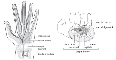 Across the base of the arch is a ligament (carpal ligament illustrated on right hand diagram) which is a little bit like an elastic band stretching between the arms of the arch and this makes the arch into a tunnel. The bones are called the carpal bones and hence the term Carpal Tunnel. Through this tunnel there are ten structures that travel from the forearm to the hand, one of which is a nerve which supplies sensation or feeling to the thumb, index finger, middle finger and half of the ring finger (as illustrated by where the median nerve goes in the diagram on the left). The conditions causing carpal tunnel generally involve thickening of the ligament at the front of the carpal tunnel. This presses down on the median nerve giving symptoms in the distribution previously stated. |
|
